DNA Microarray

A DNA microarray is a multiplex technology used in molecular biology and in medicine. It consists of an arrayed series of thousands of microscopic spots of DNA oligonucleotides, called features, each containing picomoles (10−12 moles) of a specific DNA sequence, known as probes (or reporters). This can be a short section of a gene or other DNA element that are used to hybridize a cDNA or cRNA sample (called target) under high-stringency conditions. Probe-target hybridization is usually detected and quantified by detection of fluorophore-, silver-, or chemiluminescence-labeled targets to determine relative abundance of nucleic acid sequences in the target. Since an array can contain tens of thousands of probes, a microarray experiment can accomplish many genetic tests in parallel. Therefore arrays have dramatically accelerated many types of investigation.

In standard microarrays, the probes are attached via surface engineering to a solid surface by a covalent bond to a chemical matrix (via epoxy-silane, amino-silane, lysine, polyacrylamide or others). The solid surface can be glass or a silicon chip, in which case they are colloquially known as an Affy chip when an Affymetrix chip is used. Other microarray platforms, such as Illumina, use microscopic beads, instead of the large solid support. DNA arrays are different from other types of microarray only in that they either measure DNA or use DNA as part of its detection system.

DNA microarrays can be used to measure changes in expression levels, to detect single nucleotide polymorphisms (SNPs) , to genotype or resequence mutant genomes (see uses and types section). Microarrays also differ in fabrication, workings, accuracy, efficiency, and cost (see fabrication section). Additional factors for microarray experiments are the experimental design and the methods of analyzing the data.

Genes and Genomes

Genomic DNA is located in the cell nucleus of eukaryotes, as well as small amounts in mitochondria and chloroplasts. In prokaryotes, the DNA is held within an irregularly shaped body in the cytoplasm called the nucleoid. The genetic information in a genome is held within genes, and the complete set of this information in an organism is called its genotype. A gene is a unit of heredity and is a region of DNA that influences a particular characteristic in an organism. Genes contain an open reading frame that can be transcribed, as well as regulatory sequences such as promoters and enhancers, which control the transcription of the open reading frame. In many species, only a small fraction of the total sequence of the genome encodes protein. For example, only about 1.5% of the human genome consists of protein-coding exons, with over 50% of human DNA consisting of non-coding repetitive sequences. The reasons for the presence of so much non-coding DNA in eukaryotic genomes and the extraordinary differences in genome size, or C-value, among species represent a long-standing puzzle known as the "C-value enigma."However, DNA sequences that do not code protein may still encode functional non-coding RNA molecules, which are involved in the regulation of gene expression.
Some non-coding DNA sequences play structural roles in chromosomes. Telomeres and centromeres typically contain few genes, but are important for the function and stability of chromosomes. An abundant form of non-coding DNA in humans are pseudogenes, which are copies of genes that have been disabled by mutation. These sequences are usually just molecular fossils, although they can occasionally serve as raw genetic material for the creation of new genes through the process of gene duplication and divergence.

DNA Modifying Enzymes

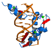
Nucleases are enzymes that cut DNA strands by catalyzing the hydrolysis of the phosphodiester bonds. Nucleases that hydrolyse nucleotides from the ends of DNA strands are called exonucleases, while endonucleases cut within strands. The most frequently used nucleases in molecular biology are the restriction endonucleases, which cut DNA at specific sequences. For instance, the EcoRV enzyme shown to the left recognizes the 6-base sequence 5′-GAT|ATC-3′ and makes a cut at the vertical line. In nature, these enzymes protect bacteria against phage infection by digesting the phage DNA when it enters the bacterial cell, acting as part of the restriction modification system. In technology, these sequence-specific nucleases are used in molecular cloning and DNA fingerprinting. Enzymes called DNA ligases can rejoin cut or broken DNA strands. Ligases are particularly important in lagging strand DNA replication, as they join together the short segments of DNA produced at the replication fork into a complete copy of the DNA template. They are also used in DNA repair and genetic recombination.

DNA Binding proteins

Structural proteins that bind DNA are well-understood examples of non-specific DNA-protein interactions. Within chromosomes, DNA is held in complexes with structural proteins. These proteins organize the DNA into a compact structure called chromatin. In eukaryotes this structure involves DNA binding to a complex of small basic proteins called histones, while in prokaryotes multiple types of proteins are involved. The histones form a disk-shaped complex called a nucleosome, which contains two complete turns of double-stranded DNA wrapped around its surface. These non-specific interactions are formed through basic residues in the histones making ionic bonds to the acidic sugar-phosphate backbone of the DNA, and are therefore largely independent of the base sequence. Chemical modifications of these basic amino acid residues include methylation, phosphorylation and acetylation. These chemical changes alter the strength of the interaction between the DNA and the histones, making the DNA more or less accessible to transcription factors and changing the rate of transcription. Other non-specific DNA-binding proteins in chromatin include the high-mobility group proteins, which bind to bent or distorted DNA.These proteins are important in bending arrays of nucleosomes and arranging them into the larger structures that make up chromosomes.

A distinct group of DNA-binding proteins are the DNA-binding proteins that specifically bind single-stranded DNA. In humans, replication protein A is the best-understood member of this family and is used in processes where the double helix is separated, including DNA replication, recombination and DNA repair. These binding proteins seem to stabilize single-stranded DNA and protect it from forming stem-loops or being degraded by nucleases.


DNA Replication

Cell division is essential for an organism to grow, but when a cell divides it must replicate the DNA in its genome so that the two daughter cells have the same genetic information as their parent. The double-stranded structure of DNA provides a simple mechanism for DNA replication. Here, the two strands are separated and then each strand's complementary DNA sequence is recreated by an enzyme called DNA polymerase. This enzyme makes the complementary strand by finding the correct base through complementary base pairing, and bonding it onto the original strand. As DNA polymerases can only extend a DNA strand in a 5′ to 3′ direction, different mechanisms are used to copy the antiparallel strands of the double helix. In this way, the base on the old strand dictates which base appears on the new strand, and the cell ends up with a perfect copy of its DNA.

Alternate DNA structures

DNA exists in many possible conformations that include A-DNA, B-DNA, and Z-DNA forms, although, only B-DNA and Z-DNA have been directly observed in functional organisms. The conformation that DNA adopts depends on the hydration level, DNA sequence, the amount and direction of supercoiling, chemical modifications of the bases, the type and concentration of metal ions, as well as the presence of polyamines in solution.

The first published reports of A-DNA X-ray diffraction patterns— and also B-DNA used analyses based on Patterson transforms that provided only a limited amount of structural information for oriented fibers of DNA. An alternate analysis was then proposed by Wilkins et al., in 1953, for the in vivo B-DNA X-ray diffraction/scattering patterns of highly hydrated DNA fibers in terms of squares of Bessel functions. In the same journal, Watson and Crick presented their molecular modeling analysis of the DNA X-ray diffraction patterns to suggest that the structure was a double-helix.

Although the `B-DNA form' is most common under the conditions found in cells, it is not a well-defined conformation but a family of related DNA conformations that occur at the high hydration levels present in living cells. Their corresponding X-ray diffraction and scattering patterns are characteristic of molecular paracrystals with a significant degree of disorder.

DNA Properties

DNA is a long polymer made from repeating units called nucleotides. The DNA chain is 22 to 26 Ångströms wide (2.2 to 2.6 nanometres), and one nucleotide unit is 3.3 Å (0.33 nm) long.Although each individual repeating unit is very small, DNA polymers can be very large molecules containing millions of nucleotides. For instance, the largest human chromosome, chromosome number 1, is approximately 220 million base pairs long.

In living organisms, DNA does not usually exist as a single molecule, but instead as a pair of molecules that are held tightly together. These two long strands entwine like vines, in the shape of a double helix. The nucleotide repeats contain both the segment of the backbone of the molecule, which holds the chain together, and a base, which interacts with the other DNA strand in the helix. A base linked to a sugar is called a nucleoside and a base linked to a sugar and one or more phosphate groups is called a nucleotide. If multiple nucleotides are linked together, as in DNA, this polymer is called a polynucleotide.

The backbone of the DNA strand is made from alternating phosphate and sugar residues.The sugar in DNA is 2-deoxyribose, which is a pentose (five-carbon) sugar. The sugars are joined together by phosphate groups that form phosphodiester bonds between the third and fifth carbon atoms of adjacent sugar rings. These asymmetric bonds mean a strand of DNA has a direction. In a double helix the direction of the nucleotides in one strand is opposite to their direction in the other strand: the strands are antiparallel. The asymmetric ends of DNA strands are called the 5′ (five prime) and 3′ (three prime) ends, with the 5' end having a terminal phosphate group and the 3' end a terminal hydroxyl group. One major difference between DNA and RNA is the sugar, with the 2-deoxyribose in DNA being replaced by the alternative pentose sugar ribose in RNA.

Genome sequencing

Full genome sequencing (FGS), also known as whole genome sequencing, complete genome sequencing, or entire genome sequencing, is a laboratory process that determines the complete DNA sequence of an organism's genome at a single time. This entails sequencing all of an organism's chromosomal DNA as well as DNA contained in the mitochondria and for plants the chloroplast as well. Almost any biological sample—even a very small amount of DNA or ancient DNA—can provide the genetic material necessary for full genome sequencing. Such samples may include saliva, epithelial cells, bone marrow, hair (as long as the hair contains a hair follicle), seeds, plant leaves, or anything else that has DNA-containing cells. Because the sequence data that is produced can be quite large (for example, there are approximately six billion base pairs in each human diploid genome), genomic data is stored electronically and requires a large amount of computing power and storage capacity. Full genome sequencing would have been nearly impossible before the advent of the microprocessor, computers, and the Information Age.

Full genome sequencing should thus not be confused with DNA profiling. The latter only determines the likelihood that genetic material came from a particular individual or group and does not contain additional information on genetic relationships, origin or suspectability on specific diseases.. It is also distinct from SNP genotyping which covers less than 0.1% of the genome. Almost all truly complete genomes are of microbes; the term "full genome" is thus sometimes used loosely to mean "greater than 95%". The remainder of this article focuses on nearly complete human genomes. In general, knowing the complete DNA sequence of an individual's genome does not, on its own, provide useful clinical information, but this may change over time as a large number of scientific studies continue to be published detailing clear associations between specific genetic variants and disease.

Genetic change

During the process of DNA replication, errors occasionally occur in the polymerization of the second strand. These errors, called mutations, can have an impact on the phenotype of an organism, especially if they occur within the protein coding sequence of a gene. Error rates are usually very low—1 error in every 10–100 million bases—due to the "proofreading" ability of DNA polymerases. (Without proofreading error rates are a thousand-fold higher; because many viruses rely on DNA and RNA polymerases that lack proofreading ability, they experience higher mutation rates.) Processes that increase the rate of changes in DNA are called mutagenic: mutagenic chemicals promote errors in DNA replication, often by interfering with the structure of base-pairing, while UV radiation induces mutations by causing damage to the DNA structure.Chemical damage to DNA occurs naturally as well, and cells use DNA repair mechanisms to repair mismatches and breaks in DNA—nevertheless, the repair sometimes fails to return the DNA to its original sequence.
In organisms that use chromosomal crossover to exchange DNA and recombine genes, errors in alignment during meiosis can also cause mutations. Errors in crossover are especially likely when similar sequences cause partner chromosomes to adopt a mistaken alignment; this makes some regions in genomes more prone to mutating in this way. These errors create large structural changes in DNA sequence—duplications, inversions or deletions of entire regions, or the accidental exchanging of whole parts between different chromosomes .

Gene regulation

The genome of a given organism contains thousands of genes, but not all these genes need to be active at any given moment. A gene is expressed when it is being transcribed into mRNA (and translated into protein), and there exist many cellular methods of controlling the expression of genes such that proteins are produced only when needed by the cell. Transcription factors are regulatory proteins that bind to the start of genes, either promoting or inhibiting the transcription of the gene. Within the genome of Escherichia coli bacteria, for example, there exists a series of genes necessary for the synthesis of the amino acid tryptophan. However, when tryptophan is already available to the cell, these genes for tryptophan synthesis are no longer needed. The presence of tryptophan directly affects the activity of the genes—tryptophan molecules bind to the tryptophan repressor (a transcription factor), changing the repressor's structure such that the repressor binds to the genes. The tryptophan repressor blocks the transcription and expression of the genes, thereby creating negative feedback regulation of the tryptophan synthesis process.
Differences in gene expression are especially clear within multicellular organisms, where cells all contain the same genome but have very different structures and behaviors due to the expression of different sets of genes. All the cells in a multicellular organism derive from a single cell, differentiating into variant cell types in response to external and intercellular signals and gradually establishing different patterns of gene expression to create different behaviors. As no single gene is responsible for the development of structures within multicellular organisms, these patterns arise from the complex interactions between many cells.

Molecule

A molecule is defined as an electrically neutral group of at least two atoms in a definite arrangement held together by very strong (covalent) chemical bonds. Molecules are distinguished from polyatomic ions in this strict sense. In organic chemistry and biochemistry, the term molecule is used less strictly and also is applied to charged organic molecules and biomolecules.

In the kinetic theory of gases, the term molecule is often used for any gaseous particle regardless of its composition. According to this definition noble gas atoms are considered molecules despite the fact that they are composed of a single non-bonded atom.

A molecule may consist of atoms of a single chemical element, as with oxygen (O2), or of different elements, as with water (H2O). Atoms and complexes connected by non-covalent bonds such as hydrogen bonds or ionic bonds are generally not considered single molecules.

Molecules as components of matter are common in organic substances (and therefore biochemistry). They also make up most of the oceans and atmosphere. A large number of familiar solid substances, however, including most of the minerals that make up the crust, mantle, and core of the Earth itself, contain many chemical bonds, but are not made of identifiable molecules. No typical molecule can be defined for ionic crystals (salts) and covalent crystals (network solids), although these are often composed of repeating unit cells that extend either in a plane (such as in graphene) or three-dimensionally (such as in diamond or sodium chloride). The theme of repeated unit-cellular-structure also holds for most condensed phases with metallic bonding. In glasses (solids that exist in a vitreous disordered state), atoms may also be held together by chemical bonds without any definable molecule, but also without any of the regularity of repeating units that characterises crystals.


Genetics

Genetics , a discipline of biology, is the science of heredity and variation in living organisms. The fact that living things inherit traits from their parents has been used since prehistoric times to improve crop plants and animals through selective breeding. However, the modern science of genetics, which seeks to understand the process of inheritance, only began with the work of Gregor Mendel in the mid-nineteenth century. Although he did not know the physical basis for heredity, Mendel observed that organisms inherit traits via discrete units of inheritance, which are now called genes.

Genes correspond to regions within DNA, a molecule composed of a chain of four different types of nucleotides—the sequence of these nucleotides is the genetic information organisms inherit. DNA naturally occurs in a double stranded form, with nucleotides on each strand complementary to each other. Each strand can act as a template for creating a new partner strand—this is the physical method for making copies of genes that can be inherited.

The sequence of nucleotides in a gene is translated by cells to produce a chain of amino acids, creating proteins—the order of amino acids in a protein corresponds to the order of nucleotides in the gene. This relationship between nucleotide sequence and amino acid sequence is known as the genetic code. The amino acids in a protein determine how it folds into a three-dimensional shape; this structure is, in turn, responsible for the protein's function. Proteins carry out almost all the functions needed for cells to live. A change to the DNA in a gene can change a protein's amino acids, changing its shape and function: this can have a dramatic effect in the cell and on the organism as a whole.

Genetic code

The genetic code is the set of rules by which a gene is translated into a functional protein. Each gene consists of a specific sequence of nucleotides encoded in a DNA (or sometimes RNA) strand; a correspondence between nucleotides, the basic building blocks of genetic material, and amino acids, the basic building blocks of proteins, must be established for genes to be successfully translated into functional proteins. Sets of three nucleotides, known as codons, each correspond to a specific amino acid or to a signal; three codons are known as "stop codons" and, instead of specifying a new amino acid, alert the translation machinery that the end of the gene has been reached. There are 64 possible codons (four possible nucleotides at each of three positions, hence 43 possible codons) and only 20 standard amino acids; hence the code is redundant and multiple codons can specify the same amino acid. The correspondence between codons and amino acids is nearly universal among all known living organisms.

Functional structure of a gene

All genes have regulatory regions in addition to regions that explicitly code for a protein or RNA product. A regulatory region shared by almost all genes is known as the promoter, which provides a position that is recognized by the transcription machinery when a gene is about to be transcribed and expressed. A gene can have more than one promoter, resulting in RNAs that differ in how far they extend in the 5' end. Although promoter regions have a consensus sequence that is the most common sequence at this position, some genes have "strong" promoters that bind the transcription machinery well, and others have "weak" promoters that bind poorly. These weak promoters usually permit a lower rate of transcription than the strong promoters, because the transcription machinery binds to them and initiates transcription less frequently. Other possible regulatory regions include enhancers, which can compensate for a weak promoter. Most regulatory regions are "upstream"—that is, before or toward the 5' end of the transcription initiation site. Eukaryotic promoter regions are much more complex and difficult to identify than prokaryoticpromoters.

Many prokaryotic genes are organized into operons, or groups of genes whose products have related functions and which are transcribed as a unit. By contrast, eukaryotic genes are transcribed only one at a time, but may include long stretches of DNA called introns which are transcribed but never translated into protein (they are spliced out before translation). Splicing can also occur in prokaryotic genes, but is less common than in eukaryotes.

Gene

A gene is a unit of heredity in a living organism. It is normally a stretch of DNA that codes for a type of protein or for an RNA chain that has a function in the organism. All proteins and functional RNA chains are specified by genes. All living things depend on genes. Genes hold the information to build and maintain an organism's cells and pass genetic traits to offspring. A modern working definition of a gene is "a locatable region of genomic sequence, corresponding to a unit of inheritance, which is associated with regulatory regions, transcribed regions, and or other functional sequence regions ". Colloquial usage of the term gene (e.g. "good genes, "hair color gene") may actually refer to an allele: a gene is the basic instruction, a sequence of nucleic acid (DNA or, in the case of certain viruses RNA), while an allele is one variant of that instruction.

The notion of a gene is evolving with the science of genetics, which began when Gregor Mendel noticed that biological variations are inherited from parent organisms as specific, discrete traits. The biological entity responsible for defining traits was later termed a gene, but the biological basis for inheritance remained unknown until DNA was identified as the genetic material in the 1940s. All organisms have many genes corresponding to many different biological traits, some of which are immediately visible, such as eye color or number of limbs, and some of which are not, such as blood type or increased risk for specific diseases, or the thousands of basic biochemical processes that comprise life.


Nucleic acid design

Nucleic acid design is the process of generating a set of nucleic acid base sequences that will associate into a desired conformation. Nucleic acid is central to the fields of DNA nanotechnology and DNA computing. It is necessary because there are many possible sequences of nucleic acid strands that will fold into a given secondary structure, but many of these sequences will have undesired additional interactions which must be avoided. In addition, there are many tertiary structure considerations which affect the choice of a secondary structure for a given design.

Nucleic acid design has similar goals to protein design: in both, the sequence of monomers is designed to favor the desired folded or associated structure and to disfavor alternate structures. However, nucleic acid design has the advantage of being a much computationally simpler problem, since the simplicity of Watson-Crick base pairing rules leads to simple heuristic methods which yield experimentally robust designs. Computational models for protein folding require tertiary structure information whereas nucleic acid design can operate largely on the level of secondary structure. However, nucleic acid structures are less versatile than proteins in their functionality.

Nucleic acid design can be considered the inverse of nucleic acid structure prediction. In structure prediction, the structure is determined from a known sequence, while in nucleic acid design, a sequence is generated which will form a desired structure.

Nucleic acid thermodynamics

Hybridization is the process of complementary base pairs binding to form a double helix. Melting is the process by which the interactions between the strands of the double helix are broken, separating the two nucleic acid strands. These bonds are weak, easily separated by gentle heating, enzymes, or physical force. Melting occurs preferentially at certain points in the nucleic acid. T and A rich sequences are more easily melted than C and G rich regions. Particular base steps are also susceptible to DNA melting, particularly T A and T G base steps. These mechanical features are reflected by the use of sequences such as TATAA at the start of many genes to assist RNA polymerase in melting the DNA for transcription.

Strand separation by gentle heating, as used in PCR, is simple providing the molecules have fewer than about 10,000 base pairs (10 kilobase pairs, or 10 kbp). The intertwining of the DNA strands makes long segments difficult to separate. The cell avoids this problem by allowing its DNA-melting enzymes (helicases) to work concurrently with topoisomerases, which can chemically cleave the phosphate backbone of one of the strands so that it can swivel around the other. Helicases unwind the strands to facilitate the advance of sequence-reading enzymes such as DNA polymerase.


DNA structure and function

It is not always the case that the structure of a molecule is easy to relate to its function. What makes the structure of DNA so obviously related to its function was described modestly at the end of the article: "It has not escaped our notice that the specific pairing we have postulated immediately suggests a possible copying mechanism for the genetic material".

The "specific pairing" is a key feature of the Watson and Crick model of DNA, the pairing of nucleotide subunits. In DNA, the amount of guanine is equal to cytosine and the amount of adenine is equal to thymine. The A:T and C:G pairs are structurally similar. In particular, the length of each base pair is the same and they fit equally between the two phosphate backbones . The base pairs are held together by hydrogen bonds, a type of chemical attraction that is easy to break and easy to reform. After realizing the structural similarity of the A:T and C:G pairs, Watson and Crick soon produced their double helix model of DNA with the hydrogen bonds at the core of the helix providing a way to unzip the two complementary strands for easy replication: the last key requirement for a likely model of the genetic molecule.

Indeed, the base-pairing did suggest a way to copy a DNA molecule. Just pull apart the two phosphate backbones, each with its hydrogen bonded A, T, G, and C components. Each strand could then be used as a template for assembly of a new base-pair complementary strand.


Paracrystalline lattice models of B-DNA structures

A paracrystalline lattice, or paracrystal, is a molecular or atomic lattice with significant amounts (e.g., larger than a few percent) of partial disordering of molecular arranegements. Limiting cases of the paracrystal model are nanostructures, such as glasses, liquids, etc., that may possess only local ordering and no global order. A simple example of a paracrystalline lattice is shown in the following figure for a silica glass: Liquid crystals also have paracrystalline rather than crystalline structures.

Highly hydrated B-DNA occurs naturally in living cells in such a paracrystalline state, which is a dynamic one in spite of the relatively rigid DNA double-helix stabilized by parallel hydrogen bonds between the nucleotide base-pairs in the two complementary, helical DNA chains (see figures). For simplicity most DNA molecular models ommit both water and ions dynamically bound to B-DNA, and are thus less useful for understanding the dynamic behaviors of B-DNA in vivo. The physical and mathematical analysis of X-ray and spectroscopic data for paracrystalline B-DNA is therefore much more complicated than that of crystalline, A-DNA X-ray diffraction patterns. The paracrystal model is also important for DNA technological applications such as DNA nanotechnology. Novel techniques that combine X-ray diffraction of DNA with X-ray microscopy in hydrated living cells are now also being developed.

DNA structure

The structure of DNA shows a variety of forms, both double-stranded and single-stranded. The mechanical properties of DNA, which are directly related to its structure, are a significant problem for cells. Every process which binds or reads DNA is able to use or modify the mechanical properties of DNA for purposes of recognition, packaging and modification. The extreme length (a chromosome may contain a 10 cm long DNA strand), relative rigidity and helical structure of DNA has led to the evolution of histones and of enzymes such as topoisomerases and helicases to manage a cell's DNA. The properties of DNA are closely related to its molecular structure and sequence, particularly the weakness of the hydrogen bonds and electronic interactions that hold strands of DNA together compared to the strength of the bonds within each strand.

Experimental techniques which can directly measure the mechanical properties of DNA are relatively new, and high-resolution visualization in solution is often difficult. Nevertheless, scientists have uncovered large amount of data on the mechanical properties of this polymer, and the implications of DNA's mechanical properties on cellular processes is a topic of active current research.

The DNA found in many cells can be macroscopic in length - a few centimetres long for each human chromosome. Consequently, cells must compact or "package" DNA to carry it within them. In eukaryotes this is carried by spool-like proteins known as histones, around which DNA winds. It is the further compaction of this DNA-protein complex which produces the well known mitotic eukaryotic chromosomes.


Molecular models of DNA

Molecular models of DNA structures are representations of the molecular geometry and topology of Deoxyribonucleic acid (DNA) molecules using one of several means, with the aim of simplifying and presenting the essential, physical and chemical, properties of DNA molecular structures either in vivo or in vitro. These representations include closely packed spheres (CPK models) made of plastic, metal wires for 'skeletal models', graphic computations and animations by computers, artistic rendering. Computer molecular models also allow animations and molecular dynamics simulations that are very important for understanding how DNA functions in vivo.

The more advanced, computer-based molecular models of DNA involve molecular dynamics simulations as well as quantum mechanical computations of vibro-rotations, delocalized molecular orbitals (MOs), electric dipole moments, hydrogen-bonding, and so on. DNA molecular dynamics modeling involves simulations of DNA molecular geometry and topology changes with time as a result of both intra- and inter- molecular interactions of DNA. Whereas molecular models of Deoxyribonucleic acid (DNA) molecules such as closely packed spheres (CPK models) made of plastic or metal wires for 'skeletal models' are useful representations of static DNA structures, their usefulness is very limited for representing complex DNA dynamics. Computer molecular modeling allows both animations and molecular dynamics simulations that are very important for understanding how DNA functions in vivo.

Nucleic acid analogues

Nucleic acid analogues are compounds structurally similar (analog) to naturally occurring RNA and DNA, used in medicine and in molecular biology research. Nucleic acids are chains of nucleotides, which are composed of three parts: a phosphate backbone, a pucker-shaped pentose sugar, either ribose or deoxyribose, and one of four nucleobases. An analogue may have any of these altered. Typically the analogue nucleobases confer, among other things, different base pairing and base stacking proprieties. Examples include universal bases, which can pair with all four canon bases, and phosphate-sugar backbone analogues such as PNA, which affect the properties of the chain (PNA can even form a triple helix).

Artificial nucleic acids include peptide nucleic acid (PNA), Morpholino and locked nucleic acid (LNA), as well as glycol nucleic acid (GNA) and threose nucleic acid (TNA). Each of these is distinguished from naturally-occurring DNA or RNA by changes to the backbone of the molecule.